Learning to love Lucilia
6th September 2024
Maggots can clean wounds and stimulate them to heal like no other treatment. Tom Ireland explores how they do it, and how to overcome the ‘yuck’ factor
Professor Yamni Nigam is passionate about maggots. Originally a parasitologist and entomologist, she now heads a group at Swansea University exploring the medicinal maggot, Lucilia sericata, and the amazing molecules that help fly larvae clean and heal chronic and infected wounds.
“We’ve published lots of papers on the molecules that we’ve discovered, and on the antibacterial, antifungal and wound-healing activity,” Nigam says. “With chronic wounds that aren’t healing, that are infected, that are smelly, that are painful, maggots can do in four days what years of other wound dressings just haven’t been able to.”
Not everyone shares her love for maggots, however. Despite several clinical trials showing maggot or ‘larval’ therapy can be more effective than standard treatments, she says patients and clinicians are still reluctant to use them. She recalls a conversation with a plastic surgeon at a conference. “I asked if he’d ever used maggots on burns. He said, ‘yes, they worked really well. But maggots? Eeeuww!’” Nigam also says that as recently as 2016, a reviewer on a crucial grant to fund her research rejected her application simply because “no-one will want to use maggots”.
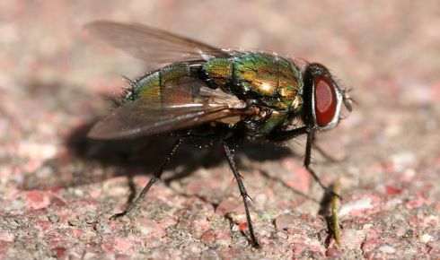 Maggot therapy is commonly carried out with larvae of the greenbottle Lucilia sericata
Despite the “persistent public disdain” Nigam says hampers its acceptance, its use is slowly growing. This is in part because of its efficacy, but also because the rising incidence of drug-resistant wound infections means people increasingly have no other option.
Nigam’s research group at Swansea University, and scientists at Biomonde (Europe’s only producers of medical-grade maggots), are both trying to understand how maggots clean wounds and seem able to stimulate tissue repair in a way that other treatments don’t. Alongside her research, Nigam runs the ‘Love a Maggot’ campaign, to try to help change people’s perception of fly larvae.
A long history
Evidence suggests that many cultures around the world have used larvae medicinally over the last thousand years – including the aboriginal Ngemba tribe of New South Wales, the Hill people of northern Myanmar, and Mayan healers in Central America. References to myiasis (wounds infected with larvae) can be found even earlier, including in the Old Testament of the Bible – ‘My body is clothed with worms and scabs, my skin is broken and festering’ (Job 7:5).
The French surgeon Ambroise Paré was the first western doctor to note the beneficial effect of fly larvae in wounds, in the 1500s. Another French surgeon, Baron Dominique-Jean Larrey, observed in the 18th and 19th century that maggots of a certain “blue fly” only removed dead tissue and had a positive effect on the remaining healthy tissue. Reports of their use to treat the wounds of war date back to the battle of St Quentin in the mid-1500s and can be found throughout the Napoleonic Wars and the American Civil War.
However, by the middle of the 19th century, an understanding of microorganisms saw doctors become more reluctant to apply non-sterile matter to open wounds. As the 20th century dawned, there were few doctors still practising this form of medicine.
The First World War came before the arrival of antibiotics like penicillin, and mortality from wounds rose to an astonishing 70%. It prompted William S Baer, a military surgeon in France, to develop specific flies and bandages for treating wounds with larvae and techniques to make them sterile. He conducted trials to test how well they worked. Thanks to his work, maggot therapy boomed again during the Second World War, with more than 300 US hospitals using maggots to treat wounds between 1930 and 1940. But the widespread availability of penicillin and dozens of other antibiotics in the 1950s meant the use of maggot therapy once again fell out of fashion and was only used as a last resort in exceptional cases.
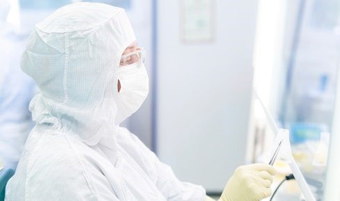 BioMonde staff even work on Christmas Day to ensure their larvae are looked after
The 1990s saw a resurgence of interest in the idea amid growing numbers of infections that would not respond to antibiotics. In the US, the physicians Ronald Sherman and Edward Pechter established a facility to create sterile fly larvae and began to promote the idea once again. In the UK, doctors John Church and Stephen Thomas founded the Biosurgical Research Unit in Bridgend in 1995, which started to produce and distribute medical maggots. This became BioMonde, which also operates manufacturing facilities in South Wales and Germany.
By the early 2000s, medicinal maggots were approved for use as ‘medical devices’ by the Food and Drug Administration in the US, and larval therapy has been available via the NHS since 2004.
The efficacy of maggot therapy is not disputed. A 2014 systematic review of 12 trials conducted since the resurgence of interest in the 1990s, including six randomised clinical trials, found that larval therapy was ‘more effective and more efficient’ at wound debridement (i.e. cleaning and removing dead tissue) when compared with conventional treatments¹. The review also found that wounds healed quicker, required fewer antibiotics, and decreased amputation risk when compared with standard treatments.
However, it remains a niche therapy, mostly used by practitioners with an interest, or as a last resort when all other treatment options have been exhausted.
The perfect maggot
Maggot therapy is most commonly carried out with larvae of the greenbottle Lucilia sericata, although other species are being investigated for potential use. The species is not only easy to rear in labs, but its larvae are physiologically incapable of breaking down living tissue. “The last thing in the world you want is for larvae to start feeding on healthy tissue around the site, or to invade the body,” says Nigam. It is thought that the larvae can’t break down a large extracellular macromolecule known as alpha-2-macroglobulin, found in living cells, restricting it to necrotic tissue only. Once they have eaten all the dead tissue, the larvae will begin to starve, rather than eat living tissue.
The larvae are voracious as they attempt to grow and moult from their first instar to their third over the course of just a few days, meaning they quickly burrow around the wound and digest as much dead tissue as they can find. This gives larval therapy the edge over traditional ‘surgical debridement’, where a surgeon uses a knife to remove dead tissue from a wound.
“The surgeon has to try to judge what is healthy tissue and what isn’t – but hasn’t got microscopes for eyes,” says Daniel Morris, a research scientist and R&D project manager at BioMonde. “Whereas the larvae just secrete enzymes that selectively degrade all of the dead tissue within that wound.”
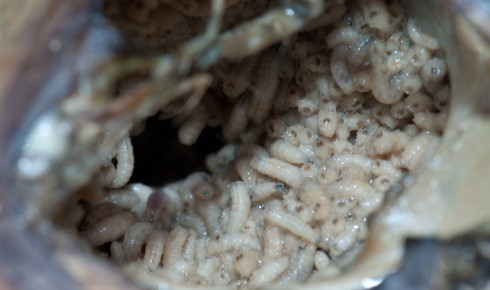 The larvae quickly burrow around the wound and digest dead tissue
Larvae then ingest the liquefied tissue. The only mouthparts they have are small hooks which help them to crawl and disturb the substrate on which they are feeding. They may push deep into the necrotic tissue but must keep their back ends exposed in order to breathe through their posterior spiracles. Their ability to crawl into unseen or hard-to-reach cavities again allows the cleaning of areas that would not be accessible with surgical debridement, but their need for oxygen limits their invasion of the body – and even means they can be considered ‘self-extracting’.
As organisms born into putrid environments, fly larvae are adept at destroying pathogenic bacteria and other microorganisms. They ingest lots of bacteria when feeding, destroying them in their gut, but also secrete powerful antibiotic compounds that further help to sterilise wounds. Research suggests some maggots have the genes to produce almost 50 different antimicrobial factors, with different ones expressed depending on the bacteria they encounter, as well as enzymes to break down tough bacterial biofilms.
“The maggot has evolved to protect itself against his own environment,” says Nigam. “And what is amazing is they can enhance their antibacterial activity if they’re in an infected wound. They can switch different genes on, depending on where they are. That’s the beauty of using a living thing in a wound; they can react to different species of bacteria, different microbes.”
The final wonder of larval therapy is that after cleaning and debriding the wound, there is good evidence that maggots actually stimulate the body’s healing process, even in wounds stuck in a chronic state of inflammation.
Research suggests that the presence of maggots helps improve oxygen perfusion to wounds and can stimulate the migration of important healing cells such as fibroblasts or microvascular endothelial cells to the wound bed². Nigam’s work has also found that growth factors expressed by maggots to move to their next larval instar are almost identical to certain human growth factors.
It’s great news for patients, but one evolutionary question springs to mind: why would fly larvae stimulate a process that closes up their home or reduces their food source?
“I don’t think the larvae intend to heal the wound,” says Micah Flores, R&D project manager at BioMonde, who also suggests that the physical disturbance caused by the maggots may also play a part in kick-starting a response from the body. “It’s just a beneficial biological coincidence.”
Making maggots
At BioMonde’s manufacturing facility in Bridgend, a team of workers keeps a colony of around 2,000 flies, in age categories by week. Some of the eggs are taken to be processed into the medicinal products, while others are used to replenish the colony. Maintaining the colony and larval products requires constant attention, with BioMonde staff even coming in on Christmas Day to provide food and water.
The flies are not kept in completely sterile conditions, as the removal of their microbiome has dismal effects on their progeny’s immunity to bacteria. The eggs, however, are thoroughly disinfected, which must be done for regulatory reasons, even though researchers believe it probably robs the larvae of the commensal microorganisms that help prime their immune system. “It’s tricky for them to go into battle without all the weapons that they would have normally,” says Flores. “You might see better results with wild maggots, but it’s the nature of a medicinal product.”
The eggs, once tested for contaminants, are then placed on nutrients to hatch in a highly controlled aseptic environment.
After hatching, the larvae are again disinfected (the female is able to pass bacteria within the egg itself, surviving the egg disinfection process). They are now ready to begin their work in wound care at hospitals and clinics across the UK and Europe.
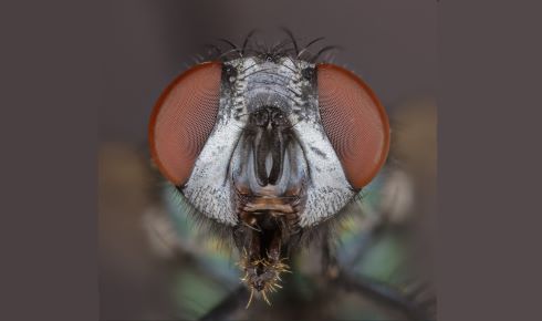 The common European greenbottle fly
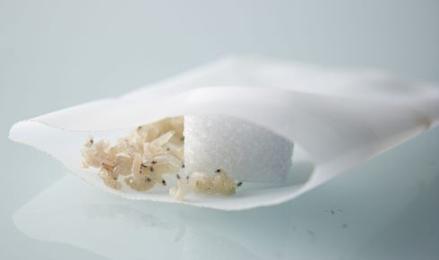
The larvae are most often packaged into a mesh dressing known as a BioBag, “sort of like a tea bag that the maggots go inside,” says Morris. The maggots are able to secrete their enzymes through the mesh and suck up the liquefied tissue, but remain contained throughout the treatment.
The larvae must be used within 48 hours of dispatch. This is one reason why larval therapy requires more thought than simply using a dressing that is stored in a cupboard: the clinician, patient and product must all arrive at the right place and time, and any delay can mean the product is ruined. (At £250 and upwards, the BioBags are far more expensive than standard dressings.) The larvae must also be kept from extremes of temperature – too much heat can cause some to speed through their larval stages and attempt to pupate without feeding.
Some patients report some pain as the maggots get to work, and the therapy can cause bleeding. Others report ‘sensations’ from the BioBag as the larvae increase in size. But mostly, the main danger is forgetting the larvae are in there and squashing them – some patients have even killed their larvae by holding their feet too close to a roaring fire. And on rare occasions, the larvae can succumb to particularly nasty bacteria in the wound.
To grow the number of people using larval therapy, there is not only the ‘yuck’ factor to overcome, but also a lack of awareness. The logistical challenges and cost can get in the way too. It all adds up to larval therapy remaining a niche treatment that too often is used as a last resort, sometimes with larvae shipped out when patients have just hours remaining before they lose a limb or develop septicaemia.
“It boggles my mind that people are still reluctant to use it early on,” says Morris. “If it’s this effective in the worst case, imagine how effective it’d be in standard cases,” adds Flores.
Hence Dr Nigam’s ‘Love a Maggot’ campaign³, launched in 2018 to counter the idea that maggots are disgusting and help people understand larval therapy. The campaign has involved hands-on activities at schools and science fairs, social science research and an interview on BBC News. BioMonde, meanwhile, brings wound care and tissue viability nurses into its facility to help show them that the BioBags are a carefully manufactured clinical product.
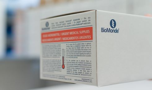 The BioBags must be used within 48 hours of dispatch, and any delay can mean the product is ruined
Maggot medicines
As well as studying how people react to maggots, Nigam is exploring the finer aspects of their secretions in detail. It could lead to new maggot-derived molecules with roles as antibiotics or in wound healing in future. For example, Nigam and colleagues have already identified one small-molecule compound, which they have called Seraticin, which is effective against numerous pathogens, including antibiotic-resistant strains⁴. Other compounds have been identified that disrupt bacterial biofilms⁵, act as potent fungicides⁶, and stimulate the formation of new epithelial cells⁷. Two enzymes, trypsin and chymotrypsin, are thought to be key in the selective debridement of dead tissue.
However exciting the discovery of these compounds is for medicine in the future, Nigam doesn’t believe there will ever be anything that can do the job like the real thing.
“I am a fan of the actual living, gorgeous little creatures, and I don’t think anything will ever replace that concoction of molecules, this adaptable moving cocktail of things for debridement, disinfection and healing.”
References
1) Sun, X. et al. A systematic review of maggot debridement therapy for chronically infected wounds and ulcers. Int. J. Infect. Dis. 25, 32–37 (2014).
2) Wang, S. et al. Maggot excretions/secretions induces human microvascular endothelial cell migration through AKT1. Mol. Biol. Rep. 37, 2719–2725 (2010).
3) Swansea University's 'Love a Maggot!' campaign. loveamaggot.com.
4) Bexfield, A. et al. The antibacterial activity against MRSA strains and other bacteria of a <500Da fraction from maggot excretions/secretions of Lucilia sericata (Diptera: Calliphoridae). Microbes Infect. 10(4),325–333 (2008).
5) Harris, L. et al. Lucilia sericata chymotrypsin disrupts protein adhesin-mediated staphylococcal biofilm formation. Appl. Environ. Microbiol. 79(4). 1393–1395 (2013).
6) Evans, R., Dudley, E., Nigam, Y. Detection and partial characterization of antifungal bioactivity from the secretions of the medicinal maggot, Lucilia sericata. Wound Repair Regen. 23(3), 361–368 (2015).
7) Bexfield, A. et al. Amino acid derivatives from Lucilia sericata excretions/secretions may contribute to the beneficial effects of maggot therapy via increased angiogenesis. Br. J. Dermatol. 162(3), 554–562 (2010).
Tom Ireland MRSB is editor of The Biologist.